By Megan Finnegan
Scientist have developed an arsenal of tools to measure brain function. For the first time, a group of psychologists and engineers have created a way to simultaneously harness the strengths of three non-invasive brain imaging tools to capture both the precise timing and location of brain activity in the human brain. The team, lead by psychology researchers Professors Florin Dolcos, Gabriele Gratton, Monica Fabiani and postdoc Matthew Moore from the University of Illinois, combined functional magnetic resonance imaging (fMRI), electroencephalography (EEG), and an imaging tool invented at the University of Illinois, called the event-related optical signal (EROS).
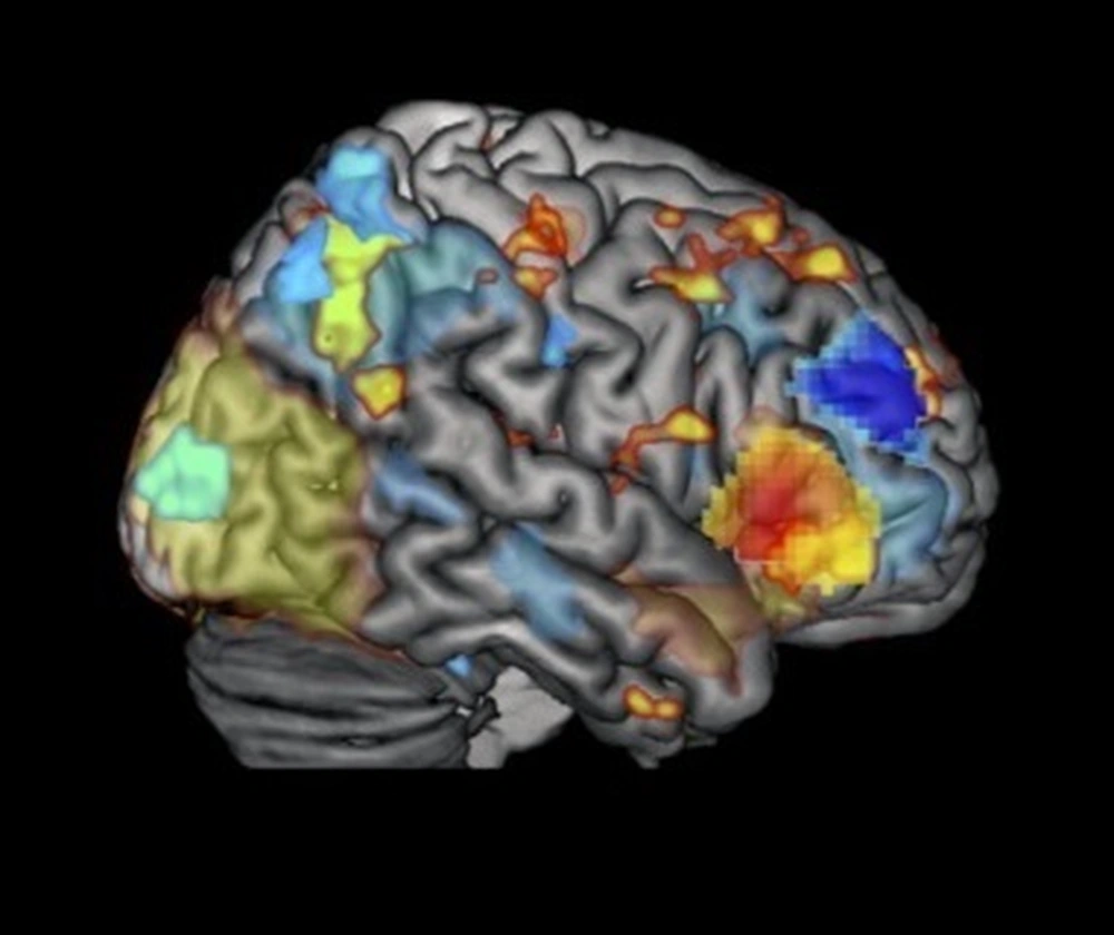
EEG and fMRI have been used to understand the link between cognitive function and neural activity for decades, but each has specific strengths and weaknesses. For example, fMRI uses strong magnetic fields to measure tiny changes in the magnetic properties of blood as active neurons deplete oxygen in the local blood supply. This allows a picture of brain activity on the level of millimeters, but with it's dependence on slow changes in blood supply, it tells us little about the timing of the activity. EEG on the other hand, relies on electrodes placed on the surface of the scalp to read electrical activity of neurons directly. It can detect changes in brain activity on the order of milliseconds, but because those signals spread across the scalp it can be difficult to localize precisely where the activity is coming from. In other words, while fMRI is good at saying where but poor at saying when brain activity is occurring, EEG is good at saying when but poor at saying where brain activity occurring. The third technique, EROS, has the high spatial resolution of fMRI and the high temporal resolution of EEG. It uses near infra-red light to penetrate the skull and measure changes in light scattering as neurons fire. Due to it's reliance on light scatter however, its measurements can only be used to capture changes occurring on the surface of the brain. However, even these surface measures allow the researchers to anchor the EEG and fMRI measures in time and space. In other words, the research team combined these three techniques such that each tool’s weakness is offset by another tool’s strengths.
This breakthrough opens the door for understanding cognition on levels of resolution that have previously been inaccessible without invasive surgical procedures. Psychologists and neuroscientists can use this approach to gain insights into how cognition unfolds across both space and time and refine theories of neural function in ways not previously feasible. Time will tell the extent to what questions will be answered, but it is no doubt that a new window of opportunity has been opened.
You can read more about this research at the Illinois News Bureau.
This research was reported in:
Moore, M., Maclin, E. L., Iordan, A. D., Katsumi, Y., Larsen, R. J., Bagshaw, A. P., Mayhew, S., Shafer, A. T., Sutton, B. P., Fabiani, M., Gratton, G., & Dolcos, F. (2021). Proof-of-concept evidence for trimodal simultaneous investigation of human brain function. Human Brain Mapping, 1– 20. https://doi.org/10.1002/hbm.25541